
Ishu Arpan, PT, PhD, Rebecca J. Willcocks, PhD, Sean C. Forbes, PhD, Richard S. Finkel, MD, Donovan J. Lott, PT, PhD, William D. Rooney, PhD, William T. Triplett, BS, Claudia R. Senesac, PT, PhD, Michael J. Daniels, ScD, Barry J. Byrne, MD, PhD, Erika L. Finanger, MD, Barry S. Russman, MD, Dah-Jyuu Wang, PhD, Gihan I. Tennekoon, MD, Glenn A. Walter, PhD, H.L. Sweeney, PhD, Krista Vandenborne, PT, PhD
Abstract
Objective
To evaluate the effects of corticosteroids on the lower extremity muscles in boys with
Duchenne muscular dystrophy (DMD) using MRI and magnetic resonance spectroscopy (MRS).
Methods
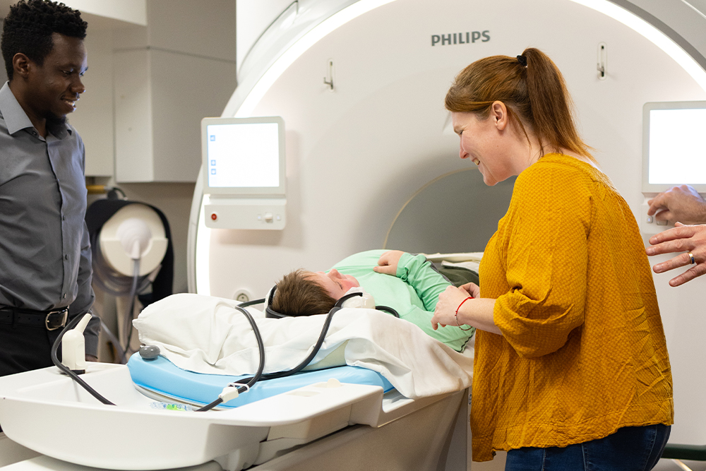
Transverse relaxation time (T2) and fat fraction were measured by MRI/MRS in lower
extremity muscles of 15 boys with DMD (age 5.0–6.9 years) taking corticosteroids and 15
corticosteroid-naive boys. Subsequently, fat fraction was measured in a subset of these boys
at 1 year. Finally, MRI/MRS data were collected from 16 corticosteroid-naive boys with DMD (age
5–8.9 years) at baseline, 3 months, and 6 months. Five boys were treated with corticosteroids
after baseline and the remaining 11 served as corticosteroid-naive controls.
Results
Cross-sectional comparisons demonstrated lower muscle T2 and less intramuscular (IM)
fat deposition in boys with DMD on corticosteroids, suggesting reduced inflammation/damage
and fat infiltration with treatment. Boys on corticosteroids demonstrated less increase in IM fat
infiltration at 1 year. Finally, T2 by MRI/MRS detected effects of corticosteroids on leg muscles
as early as 3 months after drug initiation
Conclusions
These results demonstrate the ability of MRI/MRS to detect therapeutic effects of
corticosteroids in reducing inflammatory processes in skeletal muscles of boys with DMD. Our
work highlights the potential of MRI/MRS as a biomarker in evaluating therapeutic interventions
in DMD.